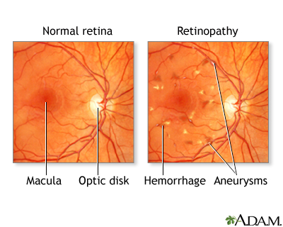
1. Diabetic Retinopathy
Nonproliferative(vascular dialation) --> proliferative(more serious, with vitreous bleeding-->floaters)
DX: fluorescein to identify vessels and laser photocoagulation to cure

2. Retinal detachment
MCC: myopia, cataracts surgery
Clinical: painless redless blurry vision, floaters, flashes, curtain coming down
Txt: lay back, laser, cryotherapy, gas injection, belt, if all above fail, surgery


3. Age-related macular degeneration
drusen
two types: a. dry, atrophic, slowly progressive blindness
b. wet, exudative, rapidly progressive blindness, need fluorescein to diagnose
Txt: a. dry: zinc, antioxidant vitamine
b. laser & verteporfin

4. Central artery occlusion
Mcc: carotid emboli, temperal arteritis, cardiac thrombi, etc.
clinical: sudden, painless, unilateral blindness, pale retina, cherry-red spot in fovea, vein box-car segmentation
DX: should undergo carotid artery image, echocardiography, thrombophilia
Txt:
a. lay back, oxygen, ocular massage,
b. acetazolamide + thrombolytics
c. anterior chamber paracentesis

5. central vein occlusion
MCC: thrombophilia
of cause glaucoma
clinical: symptoms similar to central artery occlusion, but fundoscopy is different: disk swelling, venous dilation, tortuosity,
retinal hemmorrhageno specific treatment




 Posts